
Alpha?
- informaiton
- October 24, 2017
Oshima Eye Hospital previously purchased DR-1 decade ago. We borrowed current model of DR-1 alpha, so that we compared the old and the new interferometry.
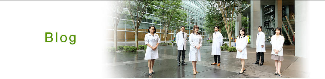
The front view of two interferometry is similar (Fig 1). The old DR-1 looks creamy color (Fig 1, right). The original color is not clear. The color may be due to the result of the passed time. The back (examiner) view is also similar (Fig 2). Current model equips several buttons on the examiner side.
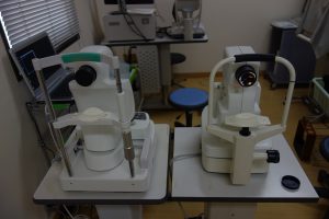
Fig 1. DR-1 (Right) and DR-1 alpha (Left)
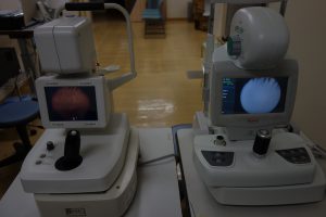
Fig 2. Examiner’s side of interferometries. DR-1 (Left) and DR-1 alpha (Right)
The interferometric image of the old DR-1 is shown as a paused image (Fig 3). The grading scale images are shown at the four edges of the images. This old model cannot record the movie thus the examiner requires to count the NIBUT during observation.
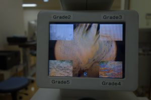
Fig 3. Observation of interferometric fringe by DR-1.
The interferometric image of the current model of DR-1 alpha is shown (Fig 4). The display is detailed and clear…it indicates passed two decades. The biggest difference of DR-1 alpha to DR-1 is the recording function of interferometric fringes. We can measure the NIBUT after obtaining movies.
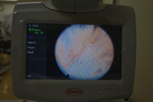
Fig 4. Observation of interferometric fringe by DR-1 alpha.
The mechanical differences are not known, but we cannot found obvious differences between two interferometry, but recording function is not replaceable. This is why current model named “Alpha”.