
What are the meibomian glands?
What are the meibomian glands?
The tear film consists of lipid layer and aqueous layer (mucin layer) (Fig. 1).
The meibomian glands are exocrine glands located in the eyelids. These glands secrete the lipid layer of the tear film and prevent the team film from evaporating.
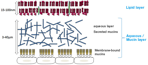
Fig. 1: Structure of the tear film
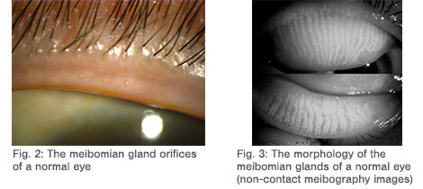
Orifices of the meibomian glands are located on the inside of the eyelids, and lipids are slowly secreted from these orifices during blinking (Fig. 2). There are about 25-30 meibomian glands in the upper eyelid and about 20-25 in the lower eyelid (Fig. 3).
A recent epidemiological study revealed that about 86% of all cases of dry eye were due to evaporative dry eye related to the meibomian glands.1
Meibomian gland dysfunction (MGD) is a condition that has garnered attention at home and abroad because of the number of people it affects.2,3
In typical clinical practice, a considerable proportion of patients visiting Ophthalmology complain of eye discomfort. This discomfort is caused by MGD, which exacerbates a patient’s quality of life and his or her quality of vision.
[References]
- 1.
- Lemp MA, Crews LA, Bron AJ, et al. Distribution of a aqueous-deficient and evaporative dry eye in a clinic based patient cohort: A retrospective study. Cornea. 31:472-8.2012
- 2.
- Amano S, Arita R et al. Meibomian Gland Dysfunction Working Group, Definition of and diagnostic criteria for meibomian gland dysfunction. Journal of the Eye, 27. 627-631, 2010.
- 3.
- Nichols KK, Foulks NG, Bron AJ, et al. The international workshop on meibomian gland dysfunction: executive summary. Invest Ophthalmol Vis Sci 52,1922-1929, 2011