Tear interferometry as a useful way to assess
the tear film lipid layer
Assessing the tear film lipid layer with tear interferometry
Tear interferometers that are currently commercially available in Japan are the LipiView® (TearScience, USA) and the DR-1α® (Kowa, Japan). The LipiView depicts the tear film lipid layer of only the bottom 1/3 of the cornea, and this device is used to determine the thickness of the lipid layer. The DR-1α depicts the tear film dynamics of the entire cornea (including its center) and it depicts the stability of the lipid layer. These devices can be used to monitor the effectiveness of treatment in patients with MGD. Clinical studies corroborating the usefulness of these devices will probably be announced in the future, and the LIME Working Group plans to present the results of similar studies.
Tear interferometry will be an area of increasing interest in the future.
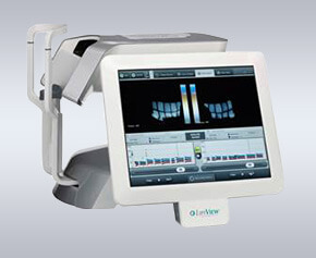
LipiView®
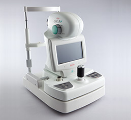
DR-1α®
The features of the DR-1α will be described below, along with materials provided by Kowa, in a layout showcasing how the device is used by the LIME Working Group.
Features of the DR-1α
- The “DR-1” came onto the market in 1997, but features such as video imaging were enhanced, and the model was fully revamped into the DR-1α in September 2015
- The DR-1α depicts optical interference in the tear film lipid layer. The device can produce still images and it is equipped with a video imaging feature that can depict the dynamics of the tear film lipid layer.
- The device can measure the non-invasive breakup time (NIBUT), and it can depict a Wide (dia.: 8 mm) or a Narrow (3.4 mm in width x 2.5 mm in height) area
- The DR-1α has a computer on-board and can be connected to files or electronic medical charts with a LAN cable.
Principle
- The lipid layer of the tear film is very thin (about 100 nm)
- When white light strikes a thin film, it reflects from the surface and the back of the film. The strength or weakness of these reflected light beams produce the phenomenon of interference
- Interference patterns change depending on the thickness of a film. Iridescence indicates a greater film thickness. Conversely, a tear film lipid layer that is spread uniformly and that has a uniform thickness will not produce an interference pattern. Instead, a monochromatic gray interference color will be visible.
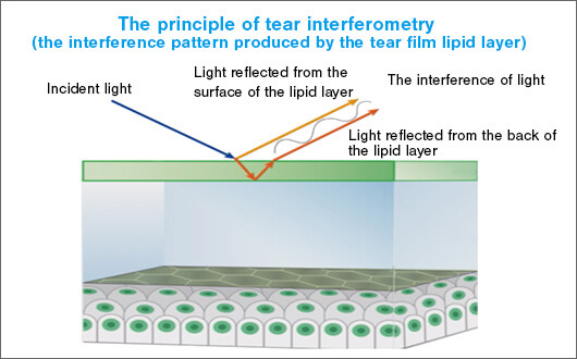
Fig. 2: Interference colors are produced by the different paths taken by light reflected by the surface of the tear film lipid layer and light reflected by the back of the lipid layer
(Slide courtesy of: Kowa ※ Partially modified from original)
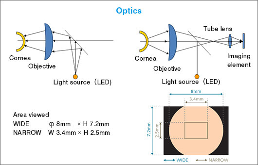
Fig. 3: Optics are designed to depict the maximum area of specular reflection from the tear film(Slide courtesy of: Kowa)
Tear interferometry allows the observation of tear film break-up in minute detail
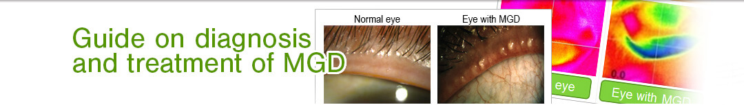
In a normal eye, the tear film is spread evenly over the cornea and there are no surface irregularities on the tear film. This results in a sharp, clean interferogram.
In a dry eye, the tear film breaks up and some of the light is scattered or reflected. This results in an inconsistent interferogram.
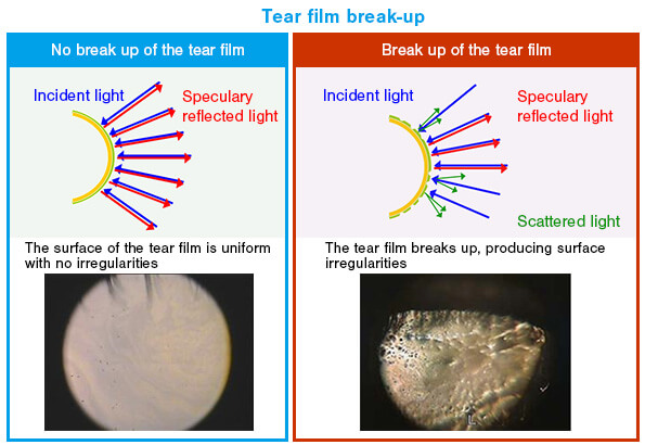
(Slide courtesy of: Kowa ※ Partially modified from original)